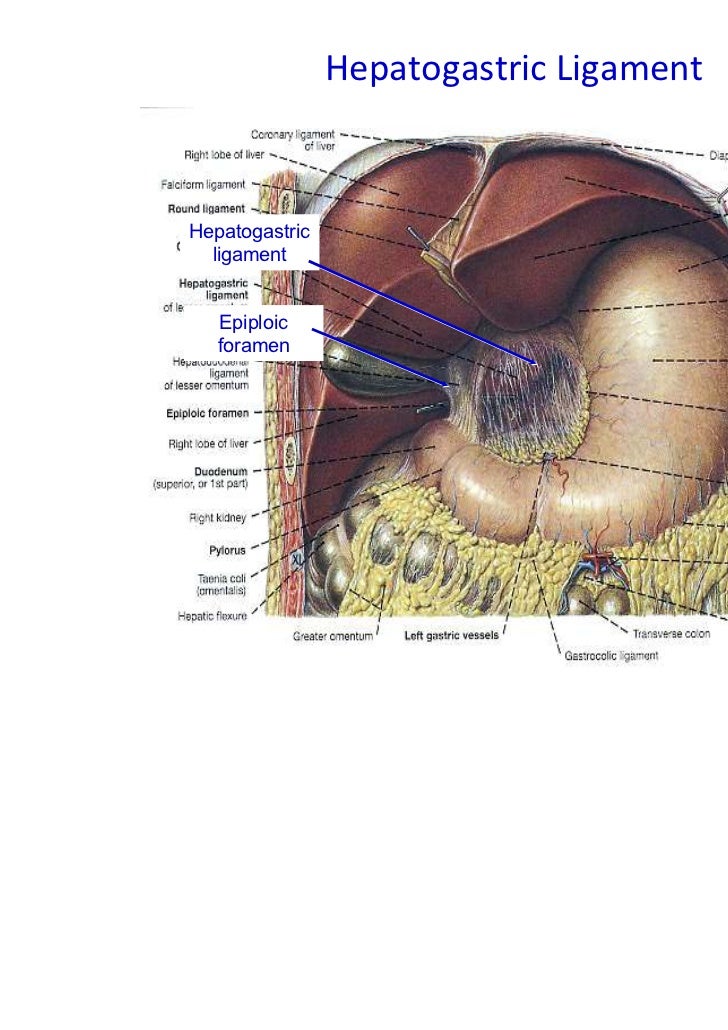
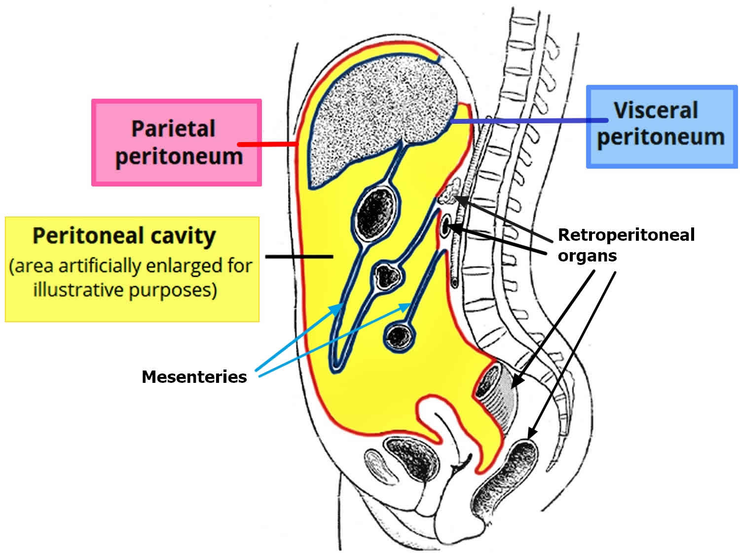
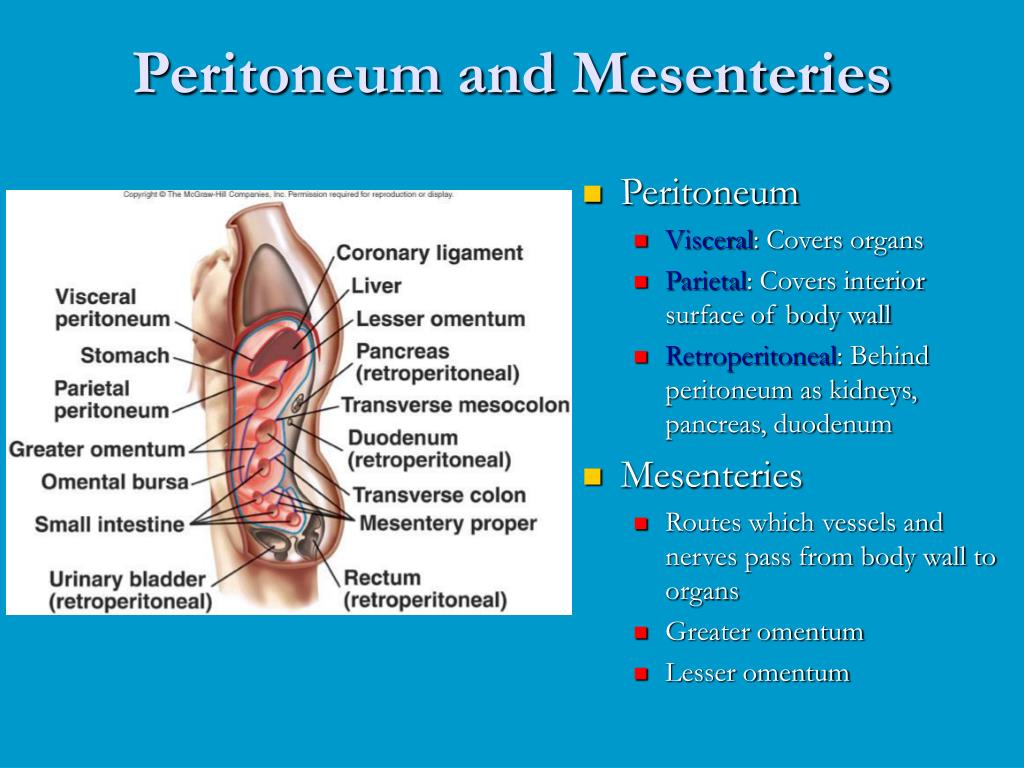
Structure Of The Peritoneum And Peritoneal Cavity Anatomy Anatomy Biology Diagrams The mesentery's classification as an organ is new. For centuries, anatomy textbooks described the mesentery as many mesenteries (plural). The medical community viewed the mesenteries as several peritoneal tissues that attach your intestines to your abdominal wall. The peritoneum is a membrane that covers your abdominal cavity and abdominal A thin layer of connective tissue is contained within the two layers of peritoneum and provides a passageway for lymphatics, nerves, arteries and veins to reach the viscera, allowing communication between the body wall and internal organs. Mesenteries are also important as they suspend or hold the organs in place to the posterior abdominal wall.. Those that are totally suspended in the cavity
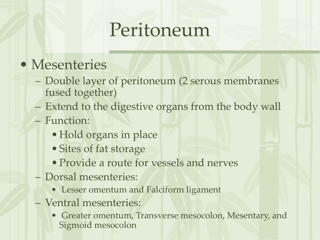
The mesentery is a connective tissue structure that is an extension of the peritoneum and connected to the intestines. The mesentery is formed of double-layer folds from part of the peritoneum Comprehensive anatomy of the peritoneum and peritoneal cavity. Organization of spaces of the greater and lesser sacs, mesenteries, and peritoneal ligaments, anchoring the organs. Characterization of the functional anatomy of the small and large bowel mesenteries. Explanation of the nerve supply of the parietal versus visceral peritoneum and the role of the visceral peritoneum in referred pain.
TeachMeAnatomy Biology Diagrams
The mesentery is a double fold of peritoneal tissue that suspends the small intestine and large intestine from the posterior abdominal wall.. It was previously thought to be a collection of discrete structures - each with separate insertions into the posterior wall. However, recent research has found the mesentery to be one contiguous structure, which has led to proposals for its
Between its two layers - parietal and visceral - is the peritoneal cavity. The peritoneum functions to support and protect abdominopelvic organs. This article will discuss the anatomy of the peritoneum, including key related topics; peritoneal cavity, omenta, mesentery, ligaments, and peritoneal relations. Viewing the peritoneal cavity Duodenum. apron-like covering that hangs in front of the small bowel and mesentery. The boundaries of the peritoneum and retroperitoneum are not usually visible on CT unless there is disease or fluid. To solidify the core anatomy, scroll through a set of images on a PACS viewer.
Mesentery: What It Is, Function, Anatomy, Location Biology Diagrams
The mesenteric model is an alternative to the peritoneal model of abdominal anatomy. It resolves several questions in abdominal anatomy, e.g., the organisation of the peritoneum 37 . The peritoneal landscape is normally described in terms of sacs, fossae, recesses, gutters, pouches and cavities 30 , 31 , 38 , 39 .
Figure 6: Development of the gut and mesenteries.A. Gut tube migrates from posterior abdominal wall, bringing the dorsal mesentery with it.B. Gut elongates and enlarges.C. Organs grow and rotate, resulting in twists in the mesenteries and peritoneal reflections (ligaments) that connect adjacent organs.